
How to read 3D molecules: caffeine, DNA and haemoglobin

Algor Lab
November 8, 2026
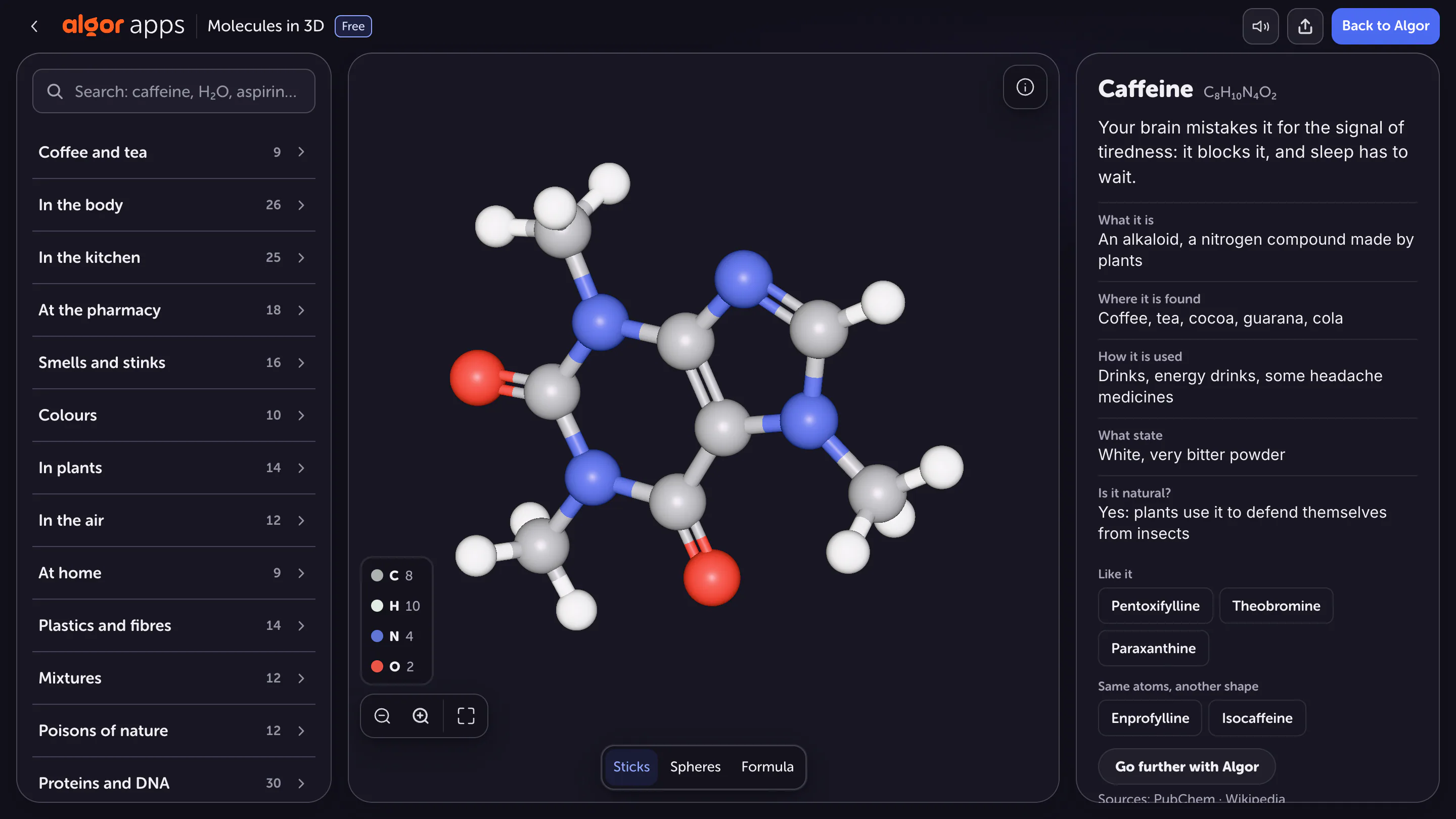
A chemical formula tells you which elements a molecule contains and in what proportions, but it does not fully describe its structure. C₈H₁₀N₄O₂ gives caffeine's composition without showing every connection or its arrangement in space. A three-dimensional model adds information, provided you understand the conventions used to display it.
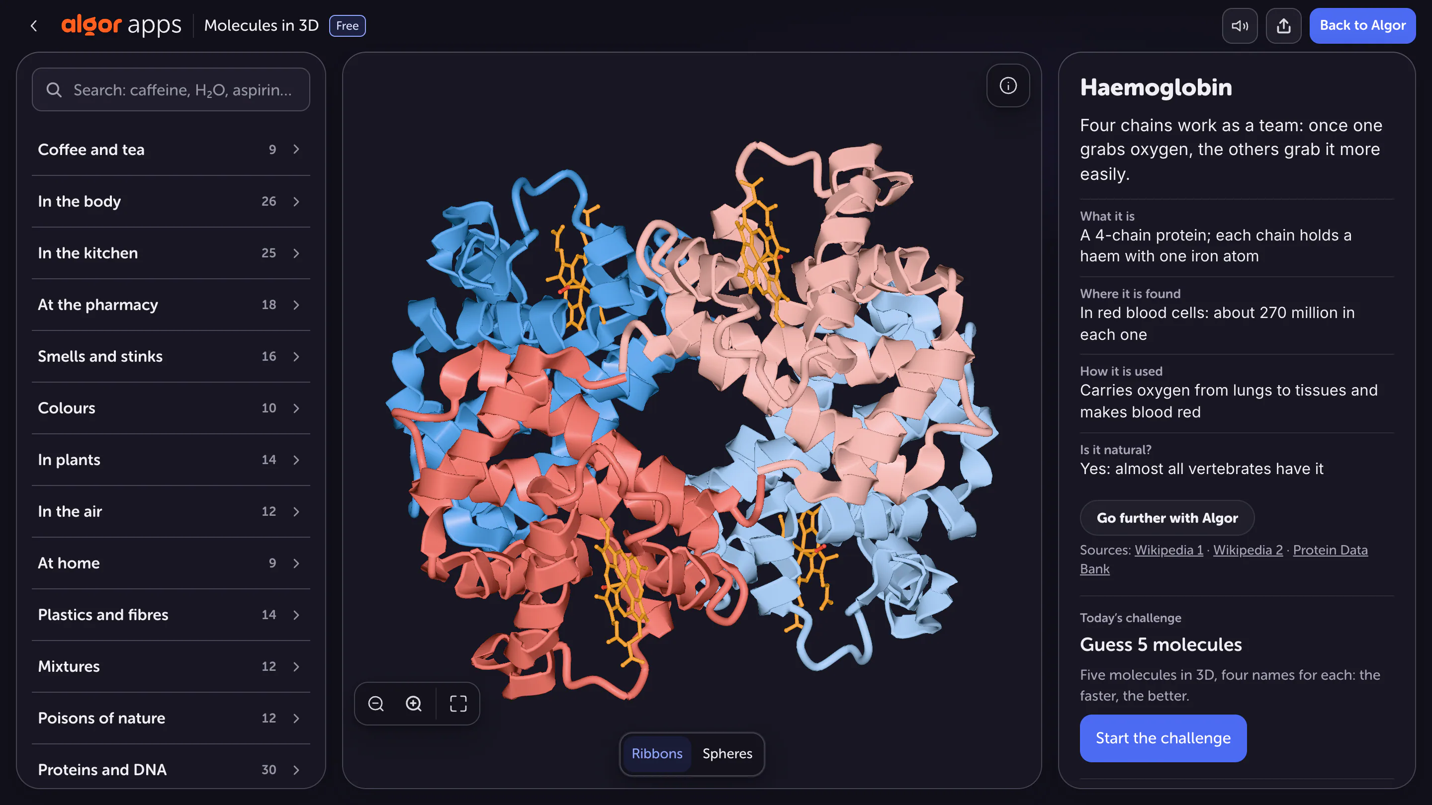
Compare three examples: a small molecule, DNA and a protein. In Algor's 3D Molecules, rotating these structures helps explain why a ball-and-stick model works well for caffeine, while ribbons and other simplified views can make much larger structures easier to read.
Read the formula before rotating the model
In caffeine's formula, the subscripts indicate eight carbon atoms, ten hydrogen atoms, four nitrogen atoms and two oxygen atoms. That gives 24 atoms per molecule, a useful first check against the atoms actually displayed. This is a molecular formula, not a map of the bonds. Molecules with the same formula can have different connections or spatial arrangements: these are examples of isomerism.
Begin with a structural question. Which atoms form rings? Where are the oxygen atoms? Which groups attach to the central structure? Looking for a specific answer is more useful than rotating continuously without recognising what changes only because of perspective. Keep the formula nearby as a check on the composition while exploring the geometry.
Balls and sticks are a visual convention
The balls represent atoms and the sticks make bonds visible. Their colours follow a graphical convention that should be checked against the available key or information. They are not the actual colours of individual atoms seen through a microscope. The sizes of the balls relative to the sticks are also chosen to make the structure readable.
Before counting atoms, check which atoms are displayed. Some representations omit hydrogens to reduce clutter. An atom hidden behind another is not missing from the molecule: rotate slowly and compare your count with the formula. A single view is not enough to establish every connection. The model is most useful when you actively test your interpretation of it.
Perspective can hide relationships
One group can appear to overlap another when you look along a particular direction. Rotating the model helps you distinguish visual overlap from a chemical bond. Two balls close together on screen are not necessarily bonded, while groups far apart in a written representation may approach each other in three-dimensional space.
Choose one atom and follow only the bonds leaving it. Describe its neighbours in words, then change the orientation and identify them again. This small exercise helps you preserve the same mental structure while the image moves. You are changing your viewpoint, not creating a new molecule each time you drag the model.
DNA requires a change of scale
For DNA, displaying every atom with equal prominence can obscure the overall organisation. A simplified representation highlights the double helix and the relationship between its strands. Those strands are not literal ribbons made from a special material: the ribbon is a way of summarising a more detailed molecular structure.
DNA strands consist of nucleotides and run in antiparallel directions in the canonical double helix. Complementary bases pair inside, while the sugar–phosphate backbones lie on the outside. DNA is a nucleic acid, not a protein. Being listed in the same catalogue of large molecular structures does not make those molecules chemically equivalent. Identify the type of molecule before transferring an explanation from one example to another.

See haemoglobin as parts working together
The most common adult haemoglobin, HbA, contains four protein chains: two alpha and two beta. A ribbon representation helps reveal their organisation without giving every atom equal visual emphasis. Colours may distinguish the subunits, but check the convention used for the selected structure rather than assuming that a colour always has the same meaning.
Each chain is associated with an iron-containing haem group involved in binding oxygen. Oxygen transport depends on more than the presence of iron: the protein's organisation and interactions between its subunits matter too. A static structure reveals components and arrangement, but it does not by itself demonstrate every movement or interaction that occurs during the molecule's activity.
Match the representation to your question
To count atoms or follow local connections, choose a view that displays those details. To recognise the folding of a large protein, ribbons may be clearer. Questions about occupied volume or an accessible surface require representations suited to those features. No single view automatically answers every question about a molecule.
Make a one-page study guide with three columns: molecule, information needed and chosen representation. Add one limitation of each view. This makes you justify the choice and separate what you can observe from what you know through theory. It also prevents a visually impressive image from becoming evidence for a claim it does not actually show.
Check your understanding without the model
Explain why caffeine's molecular formula does not describe every bond, why DNA is not a protein, and how the haem group participates in haemoglobin's function. Reopen the structures and locate the features used in your explanation. If a feature is absent from the selected display, identify the additional information you need.
The goal is to move from formula to structure and from structure to function, stating which information the model makes visible at each step. That turns a rotating image into a tool for chemical reasoning rather than an illustration you recognise but cannot explain.
Sources and further reading


Faq
Frequently Asked Questions

Related articles
Get Started with Algor
Transform your content into structured, visual and personalized learning paths.


